Ai-Hou
Wang, M.D., Ph.D.
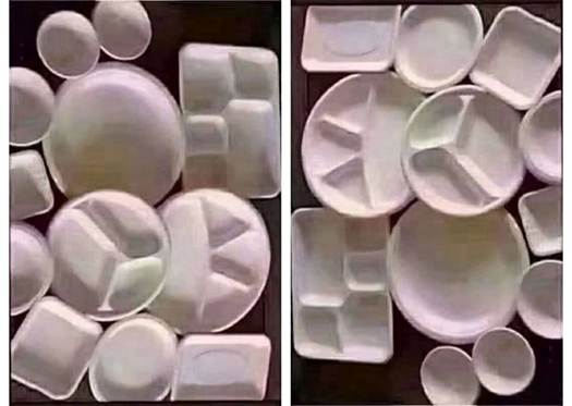
The
left and the right are the same picture, upside-down to each other.
The
depth perception is reversed.
Our
perception always assumes the illuminating light is coming from above.
And
this depth perception is quite stable.
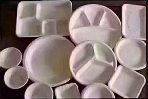
When
the image is placed horizontally, it can be viewed as light shining from the
left or from the right. The depth sensation is in a ‘bistable state’ - you
cannot see both at the same time.
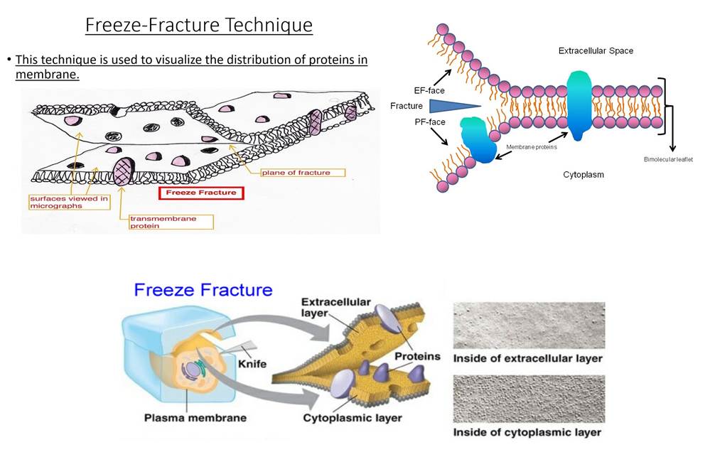
‘Freeze
fracture’ in cell biology means to separate the phospholipid bilayer of cell
membrane into inner and outer layer by freezing and splitting (see figure). You
will then be able to observe with electron microscope the interface between
inner and outer layer, to determine whether the transmembrane protein molecules
are attached to the inner layer or outer layer.
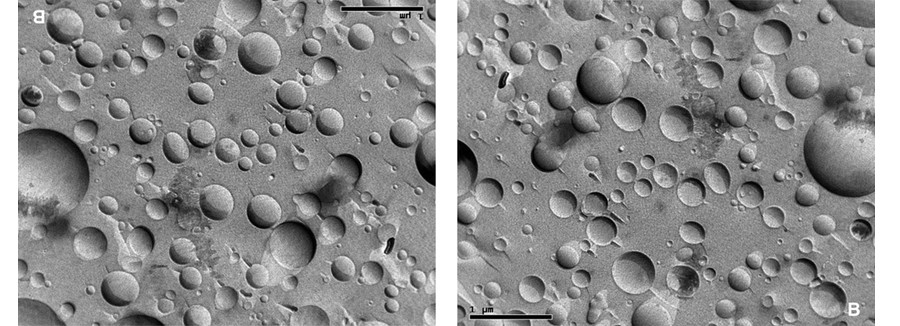
To
determine which surface of the split cell membrane are the protein molecules
attached to (see figure), we’ve got a problem:
The
left and right are the same electron microscopic picture, upside-down to each
other.
Due to
the constraint of depth perception as described above that ‘the illumination
always comes from above’, The concave hole in the left picture changes to
convex hemisphere in the right picture.
From
the left picture, the protein seems attached to the other side of the split
cell membrane; from the right picture, the protein appears attached to this
half of the split cell membrane!
This
is indeed a problem that must be solved in specimens
preparation for electron microscopy.